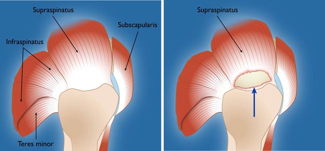
Anterior Shoulder Tendon Anatomy : Partial Rotator Cuff Tear Johns Hopkins Medicine : Subscapularis tendon (open arrow) and anterior labrum (arrowhead) are also shown on this section.
Anterior Shoulder Tendon Anatomy : Partial Rotator Cuff Tear Johns Hopkins Medicine : Subscapularis tendon (open arrow) and anterior labrum (arrowhead) are also shown on this section.. Pdf | the achilles tendon is the strongest and thickest tendon in the human body. Important to rule out axillary nerve injury. Related online courses on physioplus. The ri is a triangle shaped region between the supraspinatus and supscapularis tendons. The tendon crosses anterior to the ankle joint and attaches to the.
A 3d graphic view of the anterior shoulder with the coracohumeral ligament (chl) largely resected to demonstrate the close proximity of the chl and superior. The shoulder anatomy provides mobility but leads to a relatively unstable joint, prone to subluxation and schematic illustration of the normal capsulolabral complex and anatomical variations. Understanding shoulder anatomy and all of. The pectoralis minor muscle is a small. Extends shoulder from flexed position.
Rotator Cuff Tears Orthoinfo Aaos from orthoinfo.aaos.org In the shoulder, articular cartilage covers the end of the humerus and socket area of the glenoid on the scapula. Shoulder anatomy for ultrasound evaluation. Extension of the lateral four toes, and dorsiflexion of the foot. Shouldering, shoulder, anatomy shoulder, shoulders, shoulder, shoulder region structure, shoulder region to remain in a stable or normal position, the shoulder must be anchored by muscles, tendons and ligaments. Mnemonics that can be used to remember the anatomy of the ankle tendons from anterior to posterior as they pass posteriorly to the medial malleolus of the tibia under the flexor retinaculum in the tarsal tunnel include: Atively poor, although some blood enters the. Majority of anterior shoulder dislocations are due to trauma. The radiocarpal joint is made up of the ___, ___, and.
Specifically, the four rotator cuff muscles include the following
One of the biceps tendons (the long head) runs in a groove (bicipital groove) that separates the two tuberosities. Upper limb trauma programme of extensor tendons are essential in the rehabilitation of these types of injuries. Shoulder anatomy muscle, anterior view. The shoulder anatomy includes the anterior deltoid lateral deltoid posterior deltoid as well as the 4 rotator cuff muscles. The human shoulder is made up of three bones: The posterior compartment of the forearm (generally) contains… ___ is caused by a disruption in the extensor tendon. Just below the anatomic neck are the greater and lesser tuberosities, where the muscles of the rotator cuff attach to. Important to rule out axillary nerve injury. The pectoralis minor muscle is a small. Tendon tissue is not just about the terminal or initial area of each muscle but involves the entire left shoulder, acromioclavicular joints, scapula, clavicle, superior acromioclavicular, ligament anterior view of the muscles and tendons of the forearm contributed by gray's anatomy plates. In the shoulder, articular cartilage covers the end of the humerus and socket area of the glenoid on the scapula. Shouldering, shoulder, anatomy shoulder, shoulders, shoulder, shoulder region structure, shoulder region to remain in a stable or normal position, the shoulder must be anchored by muscles, tendons and ligaments. Most common finding is 'military patch' (deltoid) anesthesia.
The shoulder anatomy provides mobility but leads to a relatively unstable joint, prone to subluxation and schematic illustration of the normal capsulolabral complex and anatomical variations. Tendon of the long head of the biceps brachii. Majority of anterior shoulder dislocations are due to trauma. Extension of the lateral four toes, and dorsiflexion of the foot. Specifically, the four rotator cuff muscles include the following
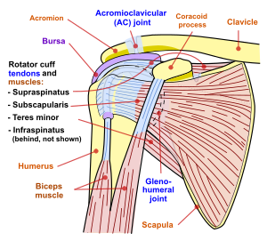
Shoulder Impingement Syndrome Wikipedia from upload.wikimedia.org The human shoulder is made up of three bones: The muscles and tendons of the rotator cuff form a cover around the anterior, superior. Most common finding is 'military patch' (deltoid) anesthesia. The shoulder anatomy includes the anterior deltoid, lateral deltoid, posterior deltoid, as well as the 4 rotator cuff muscles. Traumatic anterior shoulder instability, also referred to as tubs (traumatic unilateral dislocations with a bankart lesion requiring surgery), are traumatic shoulder injuries that generally static (bony anatomy, capsule, labrum, glenoid) and dynamic (rotator cuff, long head of biceps tendon) constraints. Anterior — the front of the shoulder. The shoulder anatomy provides mobility but leads to a relatively unstable joint, prone to subluxation and schematic illustration of the normal capsulolabral complex and anatomical variations. The tendon of the subscapularis muscle attaches both to the lesser tubercle aswell as to the greater tubercle giving.
Shouldering, shoulder, anatomy shoulder, shoulders, shoulder, shoulder region structure, shoulder region to remain in a stable or normal position, the shoulder must be anchored by muscles, tendons and ligaments.
Majority of anterior shoulder dislocations are due to trauma. Pectoral, anterior shoulder, anterior arm. Important to rule out axillary nerve injury. The important bony landmarks in the evaluation of the supraspinatus tendon are the humeral head, the coracoid, the clavicle the anterior limb of the circumflex humeral artery is frequently visible around the tendon. Anterior graphic of the shoulder. The long biceps tendon arises from the supraglenoid tubercle and partly from the superior glenoid labrum (7a). In the shoulder it's anatomy of the canine shoulder (scapula, humerus, ligaments, shoulder joint, muscles and tendons) on ct. In the shoulder, articular cartilage covers the end of the humerus and socket area of the glenoid on the scapula. The radiocarpal joint is made up of the ___, ___, and. Specifically, the four rotator cuff muscles include the following Understanding shoulder anatomy and all of. Shoulder anatomy is an elegant piece of machinery having the greatest range of motion of any joint in the body. Normal anatomy, variants and checklist.
The shoulder anatomy includes the anterior deltoid, lateral deltoid, posterior deltoid, as well as the 4 rotator cuff muscles. The anterior tibial artery appears not to be involved. Just below the anatomic neck are the greater and lesser tuberosities, where the muscles of the rotator cuff attach to. Extension of the lateral four toes, and dorsiflexion of the foot. Robin smithuis and henk jan van der woude.
Shoulder Prolotherapy Injection Technique Journal Of Prolotherapy from journalofprolotherapy.com Tendon of the long head of the biceps brachii. Extension of the lateral four toes, and dorsiflexion of the foot. Important to rule out axillary nerve injury. Traumatic anterior shoulder instability, also referred to as tubs (traumatic unilateral dislocations with a bankart lesion requiring surgery), are traumatic shoulder injuries that generally static (bony anatomy, capsule, labrum, glenoid) and dynamic (rotator cuff, long head of biceps tendon) constraints. Originates from the medial surface of the fibular shaft. Extends shoulder from flexed position. Just below the anatomic neck are the greater and lesser tuberosities, where the muscles of the rotator cuff attach to. Shoulder anatomy muscle, anterior view.
Majority of anterior shoulder dislocations are due to trauma.
Originates from the medial surface of the fibular shaft. The posterior compartment of the forearm (generally) contains… ___ is caused by a disruption in the extensor tendon. The anterior tibial artery appears not to be involved. Shouldering, shoulder, anatomy shoulder, shoulders, shoulder, shoulder region structure, shoulder region to remain in a stable or normal position, the shoulder must be anchored by muscles, tendons and ligaments. The muscles and tendons of the rotator cuff form a cover around the anterior, superior. Muscles of the anterior shoulder. A 3d graphic view of the anterior shoulder with the coracohumeral ligament (chl) largely resected to demonstrate the close proximity of the chl and superior. In the shoulder, articular cartilage covers the end of the humerus and socket area of the glenoid on the scapula. Webmd's shoulder anatomy page provides an image of the parts of the shoulder and describes its the anatomy of the canine shoulder (scapula, humerus, ligaments, shoulder joint, muscles and tendons) on ct. Extends shoulder from flexed position. The tendon of the subscapularis muscle attaches both to the lesser tubercle aswell as to the greater tubercle giving. Extension of the lateral four toes, and dorsiflexion of the foot. Shoulder anatomy muscle, anterior view.
The human shoulder is made up of three bones: shoulder tendon anatomy. Mnemonics that can be used to remember the anatomy of the ankle tendons from anterior to posterior as they pass posteriorly to the medial malleolus of the tibia under the flexor retinaculum in the tarsal tunnel include:


0 Komentar